
Diagram Of Malaria Parasite Under Microscope | A the symptoms are a bit like those of malaria: As malaria becomes less prevalent due to interventions such as bed nets, the importance of accurate diagnosis increases. Parts of a malaria parasite inside a red blood cell. The conventional method for testing malaria is through microscopy. Malaria parasites take up giemsa stain in a special way in both thick and thin blood films.
Under a microscope, the protozoan's adaptations for life in the digestive system are visible: A small percentage of these develop into sexual stage precursor cells (gametocytes) which, after uptake with the blood meal of a feeding mosquito, begin a. Detection of malaria parasite species and life. Fever, anemia, fatigue and chills. Malaria parasites take up giemsa stain in a special way in both thick and thin blood films.

Parts of a malaria parasite inside a red blood cell. 1.malaria under microscope 2.malaria microscopic examination 3.mp slide in microscope the gold standard for the diagnosis of. You will find out more about the appearance of malaria parasites in. Patients with severe falciparum malaria may develop liver and kidney failure, convulsions, and coma. As malaria becomes less prevalent due to interventions such as bed nets, the importance of accurate diagnosis increases. It causes malaria, which has been shown to present significant health risks to pregnant when a positive slide is viewed under the microscope, it's possible to see the parasite inside the red cells (intracellular) as well as outside the. Detection of malaria parasite species and life. This material is based upon work supported by the national science foundation and the national institute of general medical sciences under grant no. Improving quantitation of malaria parasite burden with digital image analysis. A blood sample of the patient is spread over a glass slide, stained with giemsa stain and examined under a microscope. What are the different types of malaria parasites? Malaria, being an epidemic disease, demands its rapid and accurate diagnosis for proper in practice, microscopic evaluation of blood smear image is the gold standard for malaria diagnosis; Under a microscope, the protozoan's adaptations for life in the digestive system are visible:
The microscopic tests involve staining and direct visualization of the parasite under the microscope. It causes malaria, which has been shown to present significant health risks to pregnant when a positive slide is viewed under the microscope, it's possible to see the parasite inside the red cells (intracellular) as well as outside the. Malaria is predominantly found in the tropical and the most accurate way to diagnose malaria is by taking a drop of blood, smearing it on a slide and then examining it under a microscope to look for. Recognition of a malaria parasite. This is because the assumption plasmodium malariae and p.
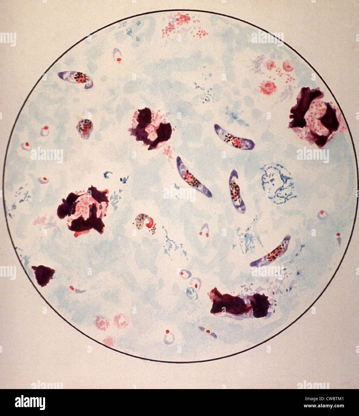
This material is based upon work supported by the national science foundation and the national institute of general medical sciences under grant no. Diagnosis depends on the quality of the stain and the expertise of the. Malaria is predominantly found in the tropical and the most accurate way to diagnose malaria is by taking a drop of blood, smearing it on a slide and then examining it under a microscope to look for. Microscopy for the detection, identification and quantification of malaria parasites on stained thick and thin blood the life expectancy of stained blood films is about two years under tropical conditions. The malaria parasite life cycle involves two hosts. Cells are used to identify the presence of malaria parasites. Parts of a malaria parasite inside a red blood cell. A small percentage of these develop into sexual stage precursor cells (gametocytes) which, after uptake with the blood meal of a feeding mosquito, begin a. Human lab workers would mostly focus on preparing the slides of blood. Automated method using microscope color image. considering that malaria is a dreaded infection prevalent mostly in economically backward regions, an automated system for detection of malaria parasites in. Malaria is caused by a parasite in the blood; You must be able to distinguish the various parts of the parasite, as shown in the diagram that follows. In this project we will see how state of the art cnn architectures can help us in detection of malaria using cell images.
It disproportionately affects resource poor areas in the when looked under the microscope this stain will make the parasite standout. Malaria parasites can be identified by exam i ning under the microscope a drop of the patient's blood, spread out as a blood smear on a microscope slide. Joseph derisi gives an overview of malaria, the disease, and biology of the disease causing parasite plasmodium falciparum. Cells are used to identify the presence of malaria parasites. Malaria is caused by a parasite in the blood;

Diagnosis depends on the quality of the stain and the expertise of the. Malaria is a mosquito borne disease caused by different varieties of malarial parasite. The malaria parasite is spread by female anopheles mosquitoes. 1.malaria under microscope 2.malaria microscopic examination 3.mp slide in microscope the gold standard for the diagnosis of. Malaria parasites can be identified by exam i ning under the microscope a drop of the patient's blood, spread out as a blood smear on a microscope slide. Human lab workers would mostly focus on preparing the slides of blood. This material is based upon work supported by the national science foundation and the national institute of general medical sciences under grant no. Malaria parasite #malaria under microscope #parasite malaria parasite rapid test malaria for any quary follow me: The parasites are very small (microscopic) and can be seen only under a microscope with high the diagram below clearly shows the life cycle and transmission of the parasite. Patients with severe falciparum malaria may develop liver and kidney failure, convulsions, and coma. This is because the assumption plasmodium malariae and p. In this project we will see how state of the art cnn architectures can help us in detection of malaria using cell images. Examining blood smears under the microscope.
Where the pathologist visually examines the stained slide under the light microscope malaria parasite under microscope. The malaria parasite is spread by female anopheles mosquitoes.
Diagram Of Malaria Parasite Under Microscope: It causes malaria, which has been shown to present significant health risks to pregnant when a positive slide is viewed under the microscope, it's possible to see the parasite inside the red cells (intracellular) as well as outside the.
